 (Image source: Gemini / Landon Phillips)
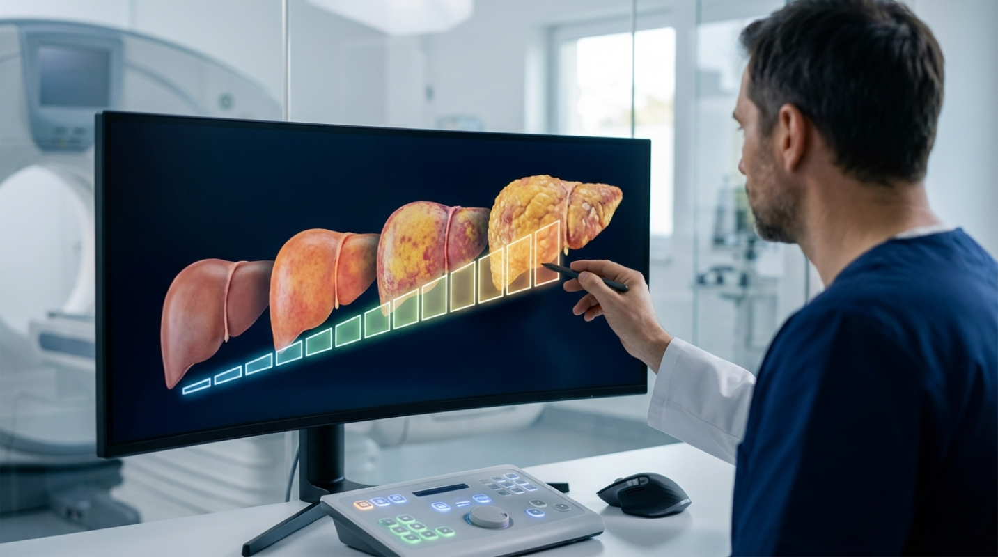
 If you’re a clinician, you know the statistics are staggering: Metabolic Dysfunction-Associated Steatotic Liver Disease (MASLD), formerly known as Non-Alcoholic Fatty Liver Disease (NAFLD), is a global epidemic. It affects up to a third of the world’s population. But having fat in your liver isn't the real problem; the spectrum of progression is.
The real danger lies in the journey from simple steatosis (fatty liver) to MASH (Metabolic Dysfunction-Associated Steatohepatitis), where inflammation begins, and finally to fibrosis (scar tissue). Fibrosis is the key predictor of long-term outcomes, determining whether a patient develops cirrhosis, liver failure, or liver cancer.
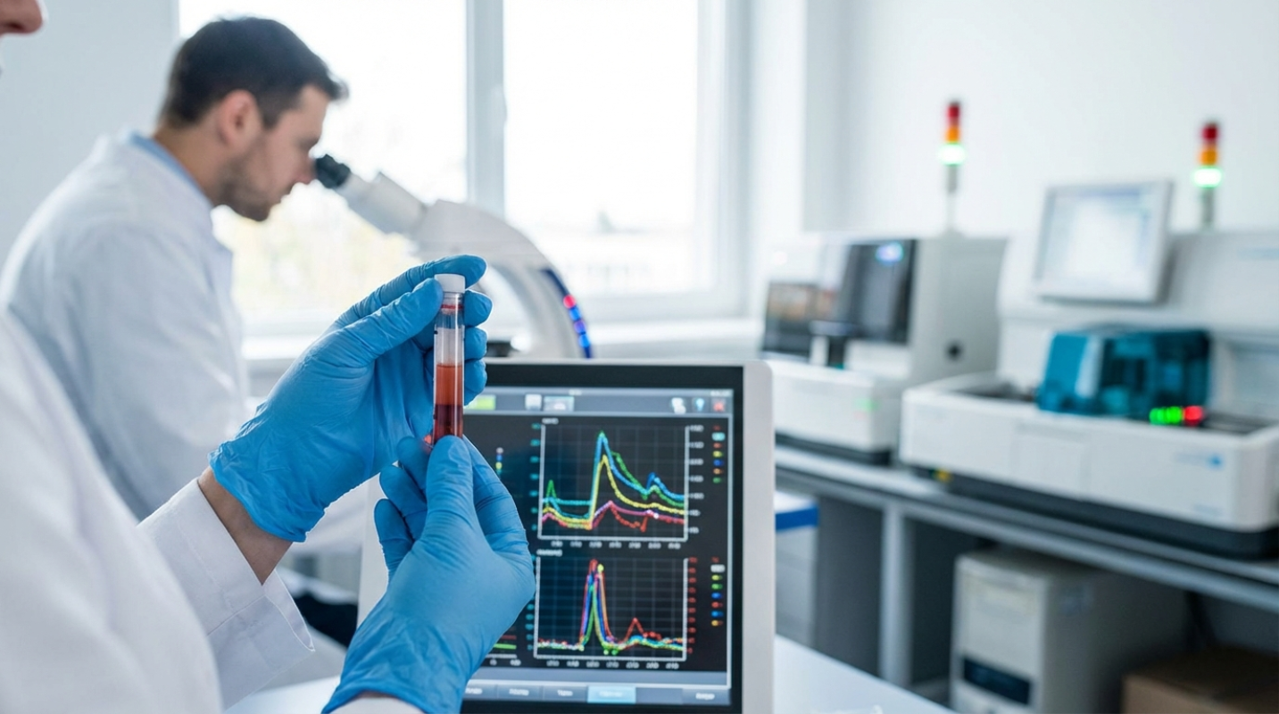
That’s why accurate staging is so important. We aren’t just looking for fat anymore; we are urgently searching for the degree of scar tissue. Historically, the liver biopsy has been the gold standard, but let’s be honest: it’s invasive, expensive, painful, and subject to significant sampling error. You’re taking a tiny piece of a large, heterogeneous organ. We need tools that are fast, repeatable, and non-invasive to manage this massive patient population. The good news is that imaging technology has delivered, moving us into an era of precision, non-invasive staging.
The Foundation: Ultrasound and Conventional MRI in Initial Assessment
When a patient walks in with elevated liver enzymes or metabolic risk factors, the first step is usually to confirm the presence of fat. That’s where standard B-mode ultrasound comes in. It’s cheap, widely available, and great for detecting moderate to severe steatosis. It tells you, qualitatively, that the liver looks "bright."
But qualitative isn't enough when you're monitoring disease progression or drug efficacy. We need numbers.
This need spurred the development of quantitative ultrasound measures, specifically the Ultrasound Attenuation Coefficient (USAC), often marketed as the Controlled Attenuation Parameter (CAP). This technology measures how much the ultrasound beam loses energy as it passes through the liver, offering a direct, numerical proxy for fat content. It’s a huge step up from simply relying on the radiologist’s subjective impression.
If you need the absolute gold standard for measuring fat, but you’re looking at Magnetic Resonance Imaging (MRI). Specifically, Proton Density Fat Fraction (PDFF). PDFF provides a precise, spatially resolved map of fat content across the entire liver. If a clinical trial demands an exact percentage of steatosis before enrollment, PDFF is the benchmark, giving you the most accurate baseline for monitoring fat reduction.
The Game Changer: Elastography Techniques for Non-Invasive Fibrosis Staging
The real clinical challenge isn't counting fat; it’s measuring stiffness—the hallmark of fibrosis. This is where elastography shines.
Transient Elastography (TE)
Transient Elastography (TE), commonly known by the brand name FibroScan, was the pioneer. It sends a mechanical pulse into the liver and measures how fast the resulting shear wave travels. Simple, fast, and highly validated. But if your patient is significantly obese or has a lot of fat (severe steatosis), the reading can be unreliable. The shear wave simply can’t penetrate or is significantly dampened.
Shear Wave and Magnetic Resonance Elastography (MRE)
Enter the next generation: Shear Wave Elastography (SWE), often integrated directly into modern ultrasound machines, and Magnetic Resonance Elastography (MRE).
MRE is arguably the most strong non-invasive measure we currently have. It uses specialized hardware to send mechanical waves through the liver while the patient is in the MRI scanner, mapping the stiffness across the entire organ.
Why is MRE so highly valued? It’s far less affected by confounding factors like obesity, ascites, or severe steatosis, which often plague ultrasound-based methods. MRE demonstrates superior accuracy, especially when diagnosing earlier stages of fibrosis. For diagnosing clinically significant fibrosis (F2 or higher), MRE achieves an impressive diagnostic accuracy (AUROC) of 0.94. Although advanced ultrasound techniques like SWE are excellent for detecting advanced fibrosis (F3/F4) with comparable accuracy, MRE is significantly more accurate for catching those important, earlier F1/F2 stages ². This higher reproducibility and accuracy make MRE the preferred tool for many high-stakes assessments.
Advanced Imaging Signatures: Functional MRI and Radiomics in Disease Progression
Although elastography tells us how stiff the liver is, it doesn't definitively tell us why—is it inflammation (MASH) or just scar tissue (fibrosis)? The next frontier involves looking beyond stiffness to capture the functional signatures of the disease.
Functional MRI techniques, such as T1 mapping or Dynamic Contrast-Improved MRI (DCE-MRI), can provide insights into inflammation and perfusion changes. These tools help assess the microcirculation and cellular environment, hinting at active inflammation that might not yet be visible as dense scar tissue.
But the most exciting development is Radiomics. Think of it like this: your standard MRI or CT scan contains thousands of quantifiable data points—texture, shape uniformity, signal intensity variation—that the human eye simply can’t process. Radiomics uses machine learning to extract these quantitative features and turn them into predictive markers.
Radiomics models are showing spectacular potential, especially when combined with elastography data. They exhibit high accuracy, with AUROC values around 0.92 to 0.93 for detecting advanced fibrosis and cirrhosis in validation cohorts ⁵. These algorithms are training themselves to predict disease progression far beyond what conventional imaging markers or expert radiologists can achieve alone. It's the digital equivalent of finding hidden patterns in the noise.
Clinical Integration and Future Directions: Precision Staging Pathways
So, with all these options, how do you decide which test to order? Current guidelines advocate for a stepwise approach that balances cost, accessibility, and accuracy. You don't jump straight to the expensive MRE for everyone ¹.
1. Initial Triage: Start with simple, inexpensive blood scores like the FIB-4 Index. This is your primary care screen to rule out advanced fibrosis.
2. Secondary Assessment: If the FIB-4 is indeterminate or high-risk, you move to an imaging-based Non-Invasive Test (NIT). This usually means VCTE/FibroScan first, due to its accessibility and lower cost.
3. Definitive Staging: MRE is reserved for cases where VCTE is inconclusive, or when precise staging of early fibrosis (F1/F2) is important, particularly for enrollment in MASH clinical trials.
Why the Biopsy Still Matters
Despite the phenomenal rise of NITs, the liver biopsy is not dead. Far from it. It remains indispensable in specific, high-stakes scenarios. It’s still the required endpoint for regulatory approval of new MASH drugs, such as the recently approved Resmetirom ³. It is the definitive tiebreaker when NIT results conflict, and it’s necessary to confirm the exact F2/F3 staging needed for initiating new targeted therapies.
Even the biopsy itself is getting an upgrade. Digital pathology and Artificial Intelligence (AI) are now analyzing biopsy slides, moving beyond subjective semi-quantitative scores to provide continuous, quantitative measures of fibrosis. This AI analysis is proving far more sensitive in detecting treatment-induced fibrosis regression than traditional histopathology ⁴.
Top Recommendations for Staging Implementation
The future of staging is moving towards automated, composite scores that combine the best of blood markers and imaging. Look for tools that integrate steatosis quantification (like CAP or PDFF) with stiffness measurements (like MRE or SWE) to identify patients who have both MASH and significant fibrosis (F2/F3)—the highest priority group for intervention.
The ultimate goal for 2026 and beyond is integrating AI and Machine Learning into these pathways. Imagine an automated algorithm that instantly combines your patient’s FIB-4 score, PDFF value, and MRE stiffness to predict their five-year risk of cirrhosis. That’s the future we’re building: personalized, precise, and completely non-invasive.
Sources:
1. Current expert consensus and clinical guidelines on MASLD staging
2. Comparative Diagnostic Accuracy of MRE vs. SWE for Early Fibrosis
3. Role of Liver Biopsy in MASH Clinical Trials (Resmetirom)
4. Digital Pathology and AI in Biopsy Analysis
5. High Diagnostic Potential of Radiomics in Staging Liver Fibrosis
This article is for informational and educational purposes only. Readers are encouraged to consult qualified professionals and verify details with official sources before making decisions. This content does not constitute professional advice.